Tuesday, May 21, 2024
Monday, May 20, 2024
شرح أهمية السرسوب للعجل الرضيع
أولاً: تعريف السرسوب:
السرسوب هو أول ما يحلب من ضرع الحيوان بعد الولادة، وهو مادة غنية بالمواد الغذائية والأجسام المناعية. يتم إنتاجه خلال الأسبوعين أو الثلاثة التي تسبق الولادة مباشرة، ويكون لونه أصفر كريمي وسميك القوام.
ثانياً: أهمية السرسوب للعجل الرضيع:
يعد السرسوب ضروريًا للعجل الرضيع لأسباب عديدة، منها:
- توفير المناعة: يولد العجل دون مناعة مكتسبة، لذلك يعتمد على السرسوب للحصول على الأجسام المناعية التي يحتاجها لمقاومة الأمراض. تحتوي جرعة واحدة من السرسوب على كمية من الأجسام المناعية تعادل ما يحتاجه العجل خلال الأسبوعين الأولين من عمره.
- تحسين وظائف الجهاز الهضمي: يساعد السرسوب على تكوين بطانة واقية في أمعاء العجل، مما يساعد على امتصاص العناصر الغذائية ومنع نمو البكتيريا الضارة.
- تسهيل عملية التبرز: يساعد السرسوب على تليين براز العجل، مما يسهل عملية التبرز ويمنع الإمساك.
- توفير الطاقة: يحتوي السرسوب على نسبة عالية من الطاقة، مما يساعد العجل على النمو والتطور.
ثالثاً: كمية السرسوب التي يحتاجها العجل:
يحتاج العجل إلى ما يعادل 10% من وزنه من السرسوب خلال أول 12 ساعة من عمره. ثم يجب أن يتناول العجل كمية من السرسوب تساوي 5% من وزنه كل 6 ساعات خلال اليومين التاليين.
رابعاً: كيفية إطعام السرسوب للعجل:
يمكن إطعام السرسوب للعجل بطرق متعددة، منها:
- الرضاعة الطبيعية: هي الطريقة المثلى لإطعام السرسوب للعجل، حيث تسمح له بالحصول على أكبر قدر من الأجسام المناعية.
- التغذية بالزجاجة: يمكن استخدام زجاجة الرضاعة لإطعام السرسوب للعجل، لكن يجب الحرص على تدفئة السرسوب إلى درجة حرارة الجسم قبل إطعام العجل.
- استخدام أنبوب التغذية: يمكن استخدام أنبوب التغذية لإطعام السرسوب للعجل إذا كان غير قادر على الرضاعة الطبيعية أو الرضاعة بالزجاجة.
خامساً: نصائح لضمان حصول العجل على الكمية الكافية من السرسوب:
- ساعد العجل على الرضاعة الطبيعية من الأم مباشرة بعد الولادة.
- إذا لم يتمكن العجل من الرضاعة الطبيعية، فقم بإطعامه السرسوب بالزجاجة أو باستخدام أنبوب التغذية.
- تأكد من تدفئة السرسوب إلى درجة حرارة الجسم قبل إطعام العجل.
- أطعم العجل كمية كافية من السرسوب في الوقت المناسب.
- راقب العجل للتأكد من أنه يتناول السرسوب بشكل صحيح.
سادساً: علامات نقص السرسوب:
- ضعف العجل.
- الإسهال أو الإمساك.
- عدم الرغبة في الرضاعة.
- الإصابة بالأمراض.
سابعاً: الوقاية من نقص السرسوب:
- تأكد من تلقي الأم جميع التطعيمات اللازمة قبل الولادة.
- وفر للعجل بيئة نظيفة وخالية من الأمراض.
- راقب العجل للتأكد من أنه يتمتع بصحة جيدة.
ثامناً: ختاماً:
يعد السرسوب ضروريًا لصحة العجل الرضيع ونموه. من خلال اتباع النصائح المذكورة أعلاه، يمكنك ضمان حصول العجل على الكمية الكافية من السرسوب للحفاظ على صحته وحمايته من الأمراض.
Saturday, May 11, 2024
Thursday, May 9, 2024
Neonatal diarrhea
to download: Neonatal Diarrhoea in Calves press here
Neonatal diarrhea, also known as calf scours, is a serious illness affecting newborn calves, particularly those within the first 28 days of life. It's a major cause of death in calves and can lead to significant economic losses for farmers.
Causes of Neonatal Diarrhea in Calves
There are two main categories of causes for neonatal diarrhea: infectious and non-infectious.
-
Infectious causes: These are caused by pathogens like bacteria, viruses, or parasites that invade the calf's intestines and disrupt its normal digestive function. Common infectious agents include:
- Bacteria: Escherichia coli (E. coli) is a major culprit, particularly enterotoxigenic E. coli (ETEC). Rotavirus, coronavirus, and Cryptosporidium are other bacterial causes.
- Viruses: Rotavirus and coronavirus are the most common viruses that cause diarrhea in calves between 5 and 15 days old.
- Parasites: Cryptosporidium is a common parasite that can cause diarrhea, especially in young calves with weak immune systems.
-
Non-infectious causes: These can be management-related issues that irritate the calf's digestive system or prevent it from absorbing nutrients properly. Examples include:
- Dietary issues: Sudden changes in milk replacer or feeding practices, overfeeding, or feeding cold milk can all contribute to scours.
- Environmental factors: Cold, drafty housing, poor hygiene, and inadequate sanitation can increase the risk of scours.
- Passive immunity failure: If a calf doesn't receive enough antibodies (immunoglobulins) from its mother's colostrum, it won't have adequate protection against infectious agents.
Signs and Symptoms of Neonatal Diarrhea in Calves
The severity of clinical signs can vary depending on the cause and severity of the diarrhea. However, some common signs include:
- Dehydration: This is the most serious consequence of scours. Calves may appear lethargic, have sunken eyes, and have dry, tacky mucous membranes.
- Diarrhea: The feces may be loose, watery, and yellow or brown in color. In some cases, there may be blood or mucus present.
- Decreased appetite: Calves may be reluctant to nurse or drink milk replacer.
- Weight loss
- Depression
- Abdominal pain
Diagnosis of Neonatal Diarrhea in Calves
A veterinarian will typically diagnose neonatal diarrhea based on the calf's history, clinical signs, and a physical examination. In some cases, they may recommend additional tests such as:
- Fecal examination: This can help identify the presence of parasites or bacteria in the feces.
- Blood tests: These can help assess the calf's hydration status and electrolyte balance.
Treatment of Neonatal Diarrhea in Calves
The treatment for neonatal diarrhea will depend on the underlying cause and the severity of the illness. However, the main goals of treatment are to:
- Correct dehydration and electrolyte imbalances: This may involve administering oral or intravenous fluids.
- Identify and address the underlying cause: This may involve antibiotics for bacterial infections, or supportive care for non-infectious causes.
- Improve gut health: Electrolytes and probiotics may be used to help restore the normal balance of bacteria in the calf's intestines.
Prevention of Neonatal Diarrhea in Calves
The best way to manage neonatal diarrhea is to prevent it from happening in the first place. Here are some key preventive measures:
- Colostrum management: Ensure newborn calves receive high-quality colostrum within the first few hours of life. Colostrum is rich in antibodies that help protect calves from infection.
- Good hygiene practices: Maintain clean calving pens and feeding equipment to reduce the spread of pathogens.
- Proper nutrition: Provide calves with a clean, fresh source of water and a milk replacer formulated for young calves. Avoid overfeeding or sudden changes in diet.
- Biosecurity measures: Isolate sick calves to prevent the spread of disease to healthy animals.
- Temperature control: Ensure calves have a warm, dry environment to help them maintain their body temperature.
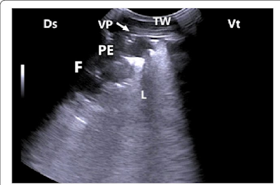
Ultrasonogram of a lung of cattle calf with fibrinous pleurisy
Ultrasonogram of a lung of cattle calf with fibrinous pleurisy exhibits interrupted visceral pleura (VP) with accumulation of fibrin (F) and inflammatory exudates forming pleural effusion (PE). TW, Thoracic wall; Ds, Dorsal; Vt, Ventral; L, Lung.
كتاب أمراض المعدية في الأبقار للأستاذ الدكتور أحمد الصوالحي
https://www.mediafire.com/file/98780g5ybcs6nbm/%2528Copy_of%2529_%25D8%25A7%25D9%2584%25D8%25A3%25D9%2585%25D8%25B1%25D8%25A7%25D8%25B6_%25D8%25A7%25D9%2584%25D9%2585%25D8%25B9%25D8%25AF%25D9%258A%25D8%25A9_%25D8%25A3%25D8%25AF.%25D9%2585%25D8%25AD%25D9%2585%25D8%25AF_%25D8%25A7%25D9%2584%25D8%25B5%25D9%2588%25D8%25A7%25D9%2584%25D8%25AD%25D9%2589_%25D8%25A7%25D9%2584%25D9%2586%25D8%25B3%25D8%25AE%25D8%25A9_%25D8%25A7%25D9%2584%25D8%25B9%25D8%25B1%25D8%25A8%25D9%258A%25D8%25A9.pdf/file
نبذة عن الكتاب:
- المؤلف: الأستاذ الدكتور أحمد الصوالحي
- التخصص: أمراض الأبقار
- المحتوى: يغطي الكتاب جميع الأمراض المعدية التي تصيب الأبقار، بما في ذلك:
- الأمراض الفيروسية: مثل داء البقري، والتهاب الفم واللسان، والحمى القلاعية
- الأمراض البكتيرية: مثل داء السل، والجمرة الخبيثة، والتهاب الأمعاء المعدي
- الأمراض الطفيلية: مثل داء الكوكسيديا، وداء البلهارسيا، وداء الديدان الشريطية
- الأمراض الفطرية: مثل داء الفطر الشعاعي، وداء الفطر الجلدي
- الجمهور المستهدف: طلاب الطب البيطري، والأطباء البيطريين، ومربي الأبقار
- اللغة: العربية
الوصول إلى الكتاب:
- نسخة ورقية: يمكنك شراء النسخة الورقية من المكتبات المتخصصة في الكتب العلمية والطبية.
- نسخة إلكترونية: قد تكون النسخة الإلكترونية متاحة على موقع المكتبة الإلكترونية لجامعة الملك فيصل بالرياض، أو على موقع دار نشر الجامعة.
مواقع ويب مفيدة:
- موقع المكتبة الإلكترونية لجامعة الملك فيصل بالرياض: https://library-web.kfupm.edu.sa/
- موقع دار نشر جامعة الملك فيصل: https://www.kfupm.edu.sa/
.ملاحظة:
- قد تختلف توفر النسخة الإلكترونية للكتاب من موقع لآخر.
- يرجى التأكد من صحة المعلومات قبل استخدامها
هل لديك أي أسئلة أخرى؟
Physiology of Domestic Animals, 3rd Edition
Physiology of Domestic Animals, 3rd Edition is a textbook designed for students of veterinary medicine, animal science, and agricultural sciences. It covers the physiology of various domestic animals, including cattle, sheep, goats, horses, pigs, dogs, cats, and poultry.
The book is divided into twenty chapters describing physiological systems such as digestion, respiration, circulation, excretion, endocrinology, reproduction, nervous system, and musculoskeletal system. Each chapter starts with a brief description of the major aspects of the topic covered.
The third edition, published in August 2016, is comprehensively revised to encompass recent progress in domestic animal physiology. It also introduces important new tools that enhance the students' learning outcome. The book is authored by Oystein V. Sjaastad, Knut Hove, and Olav Sand.
Here are some of the key features of the book:
- Emphasis on functional aspects of animal and veterinary science
- Aims at developing problem-solving skills needed for careers in veterinary medicine and animal science
- Ideal basis for university courses in general mammalian and avian physiology
- Over 600 color illustrations
- More than 3000 short, clarifying statements placed in the margins
- About 2000 questions
- Nearly 200 clinical examplesPhysiology of Domestic Animals book
https://drive.google.com/file/d/1jaUNkLI9HkJyi3WhdeIaXROTuT3Hsdwa/view
Advances in Equine Laparoscopy, 2nd Edition by Claude A. Ragle May 2024
Equine laparoscopy is a minimally invasive surgical technique that uses a laparoscope, a thin, lighted instrument, to examine the inside of a horse's abdomen. Here's a summary of its key points:
-
Diagnostic tool: Traditionally, laparoscopy has been used to diagnose abdominal problems in horses. This includes blunt or penetrating injuries, allowing vets to see internal damage without major surgery.
-
Therapeutic procedures: Recent advancements have expanded its use. Now, laparoscopy can be used for some therapeutic interventions as well. This could involve treating conditions or removing abnormalities identified during the diagnostic exam.
-
Minimally invasive: Compared to traditional exploratory surgery, laparoscopy offers several benefits. It creates smaller incisions, resulting in less pain, faster recovery times, and fewer complications for the horse.
Resources for further reading:
- If you'd like to delve deeper, the book "Advances in Equine Laparoscopy" by Claude A. Ragle is a comprehensive resource on the topic. It covers the instrumentation, basic principles, and specific techniques used in equine laparoscopy.
- https://players.brightcove.net/624142947001/experience_65fc4bc267acf1f5a45c62f3/index.html?playlistId=1794146472598325916
- https://drive.google.com/file/d/1jaUNkLI9HkJyi3WhdeIaXROTuT3Hsdwa/view?usp=sharing
Clinical Diagnostic Skills In Veterinary Surgery by Wahid Salamh Elgohul , Ahmed Ismail , Naglaa Abdelkader January 2024
- It focuses on veterinary surgery, which is a specific area of veterinary medicine dealing with surgical procedures for animals.
- Clinical diagnostic skills are highlighted, indicating the importance of diagnosing problems before surgery.
If you're interested in learning more about this specific resource, you can try searching for it online using the title and authors' names. You might find it available for purchase or through a library.
Do you have any specific questions about veterinary surgical diagnostics?
To download : https://drive.filen.io/d/6ee096dc-aec8-43ad-a132-e4ef0aece258#CHiNcJAua4KCAZTfucvjM8ff1btknqyd
Edge Shadowing Artifact
This infographic shows the result of the refraction of the ultrasound beam along the edge of a curved structure that results in a decrease in the intensity of the ultrasound beam
posterior to the curved edge and is seen as a hypoechoic zone.Follicular waves in cattle
1. The ultrasound is portable, non-invasive, provides real-time images, and produces a high-resolution characterization of the reproductive organs (Brown, (2018).
Veterinary Parasitology, 5th Edition
https://drive.google.com/file/d/1h23FVsznM3RnkkIw2Q_E7RsaF4nO3oHI/view
Content:
- Covers a wide range of parasites affecting various animals including dogs, cats, farm animals, exotics, and lab animals.
- Provides information on parasite biology, how to diagnose infections, available treatments (drugs and vaccines), and prevention methods.
Updates:
- The latest edition (presumably the fifth) reflects recent advancements in research and medical practices.
- The structure has been improved for easier access and better usability.
Importance:
- Veterinary parasitology plays a crucial role in:
- Protecting animal health and welfare
- Preventing economic losses from animal diseases
- Ensuring food safety
- Safeguarding public health (since some animal parasites can also infect humans)
- Aligning with the One Health approach, which emphasizes the interconnectedness of animal, human, and environmental health
Overall:
This textbook seems like a valuable resource for veterinarians, veterinary students, and anyone interested in animal health and the impact of parasites.
While I cannot provide PDFs directly, searching online using the title and author information you might find the textbook you described.
Wednesday, May 8, 2024
Saturday, May 4, 2024
Friday, May 3, 2024
Blackwell's Five-Minute Veterinary Consult: Small Mammal, 3rd Edition" by Barbara L. Oglesbee
Blackwell's Five-Minute Veterinary Consult: Small Mammal, 3rd Edition" by Barbara L. Oglesbee
is a veterinary resource book published in March 2024. Here's what I found about it:
- Key Features:
- Provides quick access to key information on diseases and conditions commonly affecting small exotic mammals like rabbits, rodents, ferrets, chinchillas, guinea pigs, hedgehogs, and sugar gliders.
- Concise and accessible format with consistent chapter structure for easy reference.
- Up-to-date information on diagnosis, treatment options, and common dosages.
- Includes client education handouts for better communication with pet owners.
- Benefits:
- Designed for busy veterinary practitioners, students, and anyone involved in caring for exotic companion mammals.
- Offers broad coverage of various small mammal species.
- Practical and time-saving resource for quick consultations.https://drive.google.com/file/d/1kNkzHZEpCtlFYpc0kWj8jqC4hRxZBo8x/view
Thursday, May 2, 2024
Clinical Diagnostic Skills In Veterinary Surgery by Wahid Salamh Elgohul , Ahmed Ismail , Naglaa Abdelkader January 2024
Importance of Clinical Diagnostic Skills in Veterinary Surgery:

-
Accurate Diagnosis: A core principle in veterinary surgery is having an accurate diagnosis before proceeding with an operation. This helps determine the most suitable surgical approach, predict potential complications, and assess the likelihood of success.
-
Minimally Invasive Procedures: Veterinary surgeons strive for minimally invasive techniques whenever possible. Clinical diagnostic skills aid in pinpointing the exact location and nature of the problem, allowing for targeted surgery with less damage to surrounding tissues.
-
Pre-existing Conditions: Animals undergoing surgery often have underlying health conditions. Diagnostic skills are crucial for identifying these conditions and taking necessary precautions during surgery and post-operative care.
Common Diagnostic Techniques in Veterinary Surgery:
- History Taking: A detailed medical history from the animal's owner provides valuable clues about the presenting issue and potential causes.
- Physical Examination: A thorough physical exam allows the veterinarian to assess the animal's vital signs, general health, and identify any abnormalities related to the surgical area.
- Diagnostic Imaging: X-rays, ultrasounds, CT scans, and MRIs help visualize internal structures, pinpoint the location and extent of abnormalities, and identify foreign objects.
- Laboratory Testing: Bloodwork, urinalysis, and other lab tests provide valuable information about the animal's overall health, organ function, and presence of infections.
- to download
- https://drive.filen.io/d/6ee096dc-aec8-43ad-a132-e4ef0aece258#CHiNcJAua4KCAZTfucvjM8ff1btknqyd
شروط الاضحية الواجب توافرها في عملية الذبح
بعض النصائح في عيد الأضحى المبارك أخي المواطن والمقيم كل عام وانتم بخير ١- يفضل الذبح فى المسالخ الحكومية وبإشراف الأطباء المتخصصين...
-
https://www.mediafire.com/file/98780g5ybcs6nbm/%2528Copy_of%2529_%25D8%25A7%25D9%2584%25D8%25A3%25D9%2585%25D8%25B1%25D8%25A7%25D8%25B6_%2...
-
تعتبر الإبل عرضة للإصابة بأنواع مختلفة من الطفيليات والديدان، التي تسبب تقرحات في جدار المعدة والأمعاء. الأنواع الشائعة في الإبل هى الديدا...


